Medical Imaging AI: Detect, Diagnose, Deliver
Introduction – From Film to Algorithms
The Evolution of Medical Imaging
Medical imaging has come a long way since the early days of film-based X-rays. In the past, radiologists would manually review every film, relying purely on their trained eyes to detect fractures, tumors or other signs of disease. This process, while effective, was time-consuming, highly subjective and prone to occasional human error — especially when working under pressure.
The transition to digital imaging and PACS (Picture Archiving and Communication Systems) brought enormous improvements. Images could now be stored, retrieved and shared instantly across hospital networks. But even with digital technology, a new challenge emerged: the sheer volume of imaging studies exploded. Today, radiologists must read hundreds of complex cases each day, often under tight time constraints.
The Rising Challenge for Healthcare
Several trends are putting increasing pressure on medical imaging departments around the world:
Growing Demand: Aging populations and greater access to advanced diagnostics mean more scans are being ordered than ever before.
Radiologist Shortage: Many regions face a shortage of qualified radiologists, leading to longer waiting times for results.
Complexity of Scans: Modern imaging modalities like multi-phase CT and high-resolution MRI generate massive amounts of data per patient.
Risk of Missed Diagnoses: Fatigue, time pressure and subtle findings can lead to occasional oversight — with serious consequences for patient care.
These factors make it clear that human expertise alone, no matter how skilled, needs technological support to keep up with modern healthcare demands.
Enter AI: A New Era for Radiology
Artificial Intelligence and particularly deep learning technologies, are now changing the game. Instead of relying solely on human interpretation, AI algorithms trained on millions of images can help:
Detect abnormalities faster
Highlight suspicious regions for closer review
Prioritize urgent cases for immediate attention
Standardize readings across different hospitals and clinicians
By working alongside radiologists — not replacing them — AI helps ensure that every scan gets a thorough, consistent review, even under heavy workloads.
What This Blog Post Will Explore
In this article, we’ll dive into how two powerful types of deep learning models — Convolutional Neural Networks (CNNs) and Vision Transformers (ViTs) — are transforming the detection, diagnosis and delivery of medical imaging results.
You'll learn:
Why CNNs remain a cornerstone for tasks like tumor spotting and fracture detection.
How Vision Transformers are unlocking new capabilities for subtle and complex diagnoses.
What a real-world AI imaging pipeline looks like, from scan acquisition to clinical report generation.
How hospitals and imaging centers are safely deploying AI while meeting strict privacy regulations.
AI-powered imaging is not science fiction — it’s happening today, reshaping healthcare delivery and improving patient outcomes around the globe. Let’s explore how it all comes together.
Why Deep Learning Is Reshaping Diagnostic Imaging
Data, Data, Data: The New Fuel for Diagnosis
Medical imaging produces enormous amounts of data every day. Every X-ray, CT scan, MRI and ultrasound adds to an ever-growing mountain of information. This explosion of medical images has created the perfect environment for training deep learning models.
With access to millions of labeled examples — such as scans showing tumors, fractures or other abnormalities — AI systems can learn to recognize patterns with astonishing precision.
The more high-quality, diverse data an AI system sees, the better it becomes at spotting subtle or rare conditions. This growing "data ocean" is one of the key reasons why deep learning is such a powerful fit for medical imaging today.
Cloud Computing and GPUs: Instant Scalability
Another major enabler of AI in healthcare is the rise of powerful computing hardware and cloud infrastructure.
Training deep neural networks requires significant processing power, especially when dealing with 3D scans or very high-resolution images. Thanks to cloud platforms and GPUs (graphics processing units), what once took days or weeks can now be done in a matter of hours.
Today, healthcare providers can even use ready-to-go APIs to plug powerful AI image analysis into their workflows without needing to build complex models themselves. This approach dramatically lowers the barrier to entry for hospitals, clinics and telemedicine providers.
APIs like Object Detection and Image Labelling — available in cloud-based form — allow rapid deployment of automated workflows, from triaging chest X-rays for pneumonia to flagging potential fractures in trauma scans.
Regulatory Green Light: Real-World Validation
One of the most exciting developments has been the increasing acceptance of AI by medical regulators.
In recent years, the U.S. Food and Drug Administration (FDA) and other authorities around the world have approved several AI-powered systems for clinical use. Some examples include:
AI tools that detect strokes on brain CT scans within minutes.
AI assistants that spot early signs of breast cancer in mammograms.
Lung nodule detection software for improving lung cancer screening.
These approvals show that AI is not just an academic exercise — it is delivering tangible results for real patients, under strict safety and effectiveness standards.
Better Outcomes, Faster Care, Lower Costs
Hospitals and healthcare providers are realizing that AI is not just about efficiency — it’s about better outcomes:
Faster diagnosis leads to quicker treatment, which can be life-saving in emergencies like strokes.
More consistent readings reduce the risk of missed findings and second opinions.
Prioritizing critical cases ensures that the most urgent patients are treated first.
Lower overall costs are achieved by reducing unnecessary procedures, cutting waiting times and optimizing radiologist workloads.
In value-based healthcare models — where providers are rewarded for outcomes rather than just services — integrating AI into diagnostic imaging is becoming a major competitive advantage.
The New Normal for Imaging
Deep learning has shifted the expectations for diagnostic imaging.
Today, radiologists working with AI tools can be faster, more accurate and more confident — especially when dealing with high volumes or complex cases. Patients benefit from quicker answers and better care, while healthcare systems benefit from streamlined operations.
In the next sections, we’ll look closely at the specific deep learning technologies making this revolution possible — starting with the workhorse of medical imaging AI: Convolutional Neural Networks (CNNs).
CNNs: The Proven Workhorse for Tumor and Fracture Detection
What Are Convolutional Neural Networks?
Convolutional Neural Networks (CNNs) are a type of deep learning model designed specifically for analyzing images. They work by scanning an image piece by piece — almost like looking at it through a small window — and learning to recognize important patterns, such as edges, textures and shapes.
This "local view" approach makes CNNs excellent at spotting fine details, like small fractures in bones or irregular masses in soft tissues. Over multiple layers, CNNs combine simple features into more complex structures, enabling them to identify subtle signs of disease that even experienced radiologists might miss.
Why CNNs Fit Medical Imaging So Well
Medical images are rich in detail and often, early signs of illness are very small or hidden within complex structures. CNNs are especially good at:
Recognizing local anomalies, such as tiny tumors or hairline fractures.
Handling different imaging types, like grayscale X-rays, colorful histology slides or 3D CT volumes.
Working even with noisy or imperfect images, which are common in real-world hospital settings.
Thanks to their flexibility and precision, CNNs have become the backbone of many AI applications in radiology.
Popular CNN Architectures in Healthcare
Several well-known CNN designs have proven to be very effective for medical image tasks:
U-Net: A CNN built specifically for image segmentation — marking the exact boundaries of tumors organs or lesions in scans.
ResNet: A very deep CNN that solves complex classification problems without getting "confused" as it goes deeper.
EfficientNet: A lightweight, high-performance CNN ideal for edge deployments where computing resources are limited, such as portable ultrasound machines.
These architectures have been used as the foundation for many FDA-cleared AI tools and academic breakthroughs in medical imaging.
Real-World Success Stories: How CNNs Help Detect Diseases
Here are a few practical examples of CNNs making a real difference in healthcare today:
Breast Cancer Screening
CNN-based models assist radiologists in reviewing mammograms by automatically flagging suspicious masses and micro-calcifications. This not only speeds up reading times but also reduces the chances of missing early-stage cancers.
Skeletal Fracture Detection
In emergency departments, AI models powered by CNNs can analyze wrist, hip or ankle X-rays within seconds. They highlight potential fractures so that clinicians can prioritize cases without delay — critical for avoiding complications like internal bleeding or nerve damage.
Brain Hemorrhage Triage
When it comes to stroke care, every minute counts. CNNs trained on CT brain scans can detect hemorrhages in under 45 seconds. By pushing urgent cases to the top of a radiologist’s queue, AI dramatically shortens the "door-to-needle" time for life-saving treatment.
How AI APIs Bring CNN Power to Healthcare Workflows
Healthcare providers don’t always need to build their own CNNs from scratch. Thanks to cloud-based APIs, hospitals can easily integrate advanced image analysis into their existing systems.
For example:
Object Detection APIs can automatically locate tumors or fractures in incoming scans.
Image Labelling APIs can classify images into normal vs. abnormal or flag different types of conditions.
Image Anonymization APIs ensure patient privacy is protected when sharing or processing data.
These tools allow clinics to test and deploy CNN-powered solutions quickly, without heavy upfront investments in hardware or software development.
The Bottom Line
CNNs have earned their place as the "workhorse" of medical imaging AI.
They are fast, reliable and incredibly powerful at detecting the small but critical details that can change a patient’s life. As we'll see next, a new generation of AI models — Vision Transformers — is now taking these capabilities even further, adding a new dimension to what AI can achieve in healthcare.
Vision Transformers: Long-Range Context for Subtle Anomalies
What Makes Vision Transformers Different?
While CNNs are excellent at picking up local patterns like edges and textures, they sometimes struggle with understanding the bigger picture — for example, the overall shape of an organ or how different parts of an image relate to each other.
This is where Vision Transformers (ViTs) come in.
Inspired by how models handle language (capturing relationships between words in a sentence), Vision Transformers look at an entire image as a collection of smaller patches. They use a technique called self-attention to figure out how each patch relates to every other patch.
In simple terms, ViTs are great at understanding both the fine details and the global structure of an image — making them perfect for complex medical imaging tasks.
Why Vision Transformers Matter for Healthcare
Medical images often require understanding both local features and overall patterns.
For example:
Identifying symmetry or asymmetry between two lungs on a chest X-ray.
Analyzing the spread of a liver lesion across multiple CT slices.
Detecting tiny changes across a large area, like early diabetic damage in the retina.
Vision Transformers can capture these relationships better than traditional CNNs. This makes them especially powerful for detecting subtle or complex abnormalities that might otherwise go unnoticed.
How Vision Transformers Work in Practice
Vision Transformers start by breaking an image into small, fixed-size patches — similar to cutting a photo into little squares.
Each patch is treated like a "word" in a sentence and the model learns how all patches relate to each other through self-attention mechanisms.
By doing this, ViTs can:
Focus on critical regions even if they are far apart in the image.
Model complex shapes and patterns that are important for diagnosis.
Adapt flexibly to different image sizes and types without major changes to the architecture.
Although they usually require more data and computational power to train than CNNs, their ability to grasp complex spatial relationships opens up exciting new possibilities for medical diagnostics.
Early Wins: Real-World Applications of Vision Transformers
Vision Transformers are already making their mark in medical imaging. Here are a few notable examples:
Liver Lesion Classification
In multi-phase CT scans, it can be hard to tell benign cysts from malignant tumors because their appearance changes depending on the timing of the contrast agent. ViTs have shown strong performance in classifying liver lesions by understanding how the lesion's appearance evolves across different phases.
Retinal Disease Detection
Optical Coherence Tomography (OCT) scans produce cross-sectional images of the retina. ViTs can analyze these 3D-like structures to detect early signs of diabetic macular edema — a leading cause of vision loss — even before patients notice symptoms.
Digital Pathology on Gigapixel Slides
Analyzing whole-slide images of tissue samples is a monumental task because a single file can contain billions of pixels. Vision Transformers can process these slides patch by patch, finding cancerous regions without needing to look at every pixel manually.
These successes highlight how ViTs are pushing the boundaries of what AI can detect and diagnose.
Making Vision Transformers Practical: Model Compression
One challenge with Vision Transformers is their size — they tend to be computationally heavier than CNNs.
However, researchers and engineers have developed several ways to make them faster and more efficient for real-world use:
Pruning: Cutting out parts of the model that don't contribute much to the final decision.
Quantization: Reducing the precision of calculations to speed up processing without losing accuracy.
Knowledge Distillation: Training a smaller "student" model to mimic the behavior of a larger "teacher" model.
These techniques help deploy ViT-based models not just in big hospitals but also on cloud platforms and even at the edge — such as in portable ultrasound machines or mobile imaging apps.
Healthcare APIs powered by optimized ViTs can soon offer even more complex analysis, from detecting rare tumors to predicting disease progression based on subtle image patterns.
The Future Looks Bright
Vision Transformers are adding a new dimension to medical imaging AI.
By combining the strengths of local detail recognition and global context understanding, they help healthcare providers detect diseases earlier, make more accurate diagnoses and deliver better patient care.
In the next section, we’ll explore how everything comes together — how AI workflows go from raw medical images to actionable clinical reports that doctors can use right away.
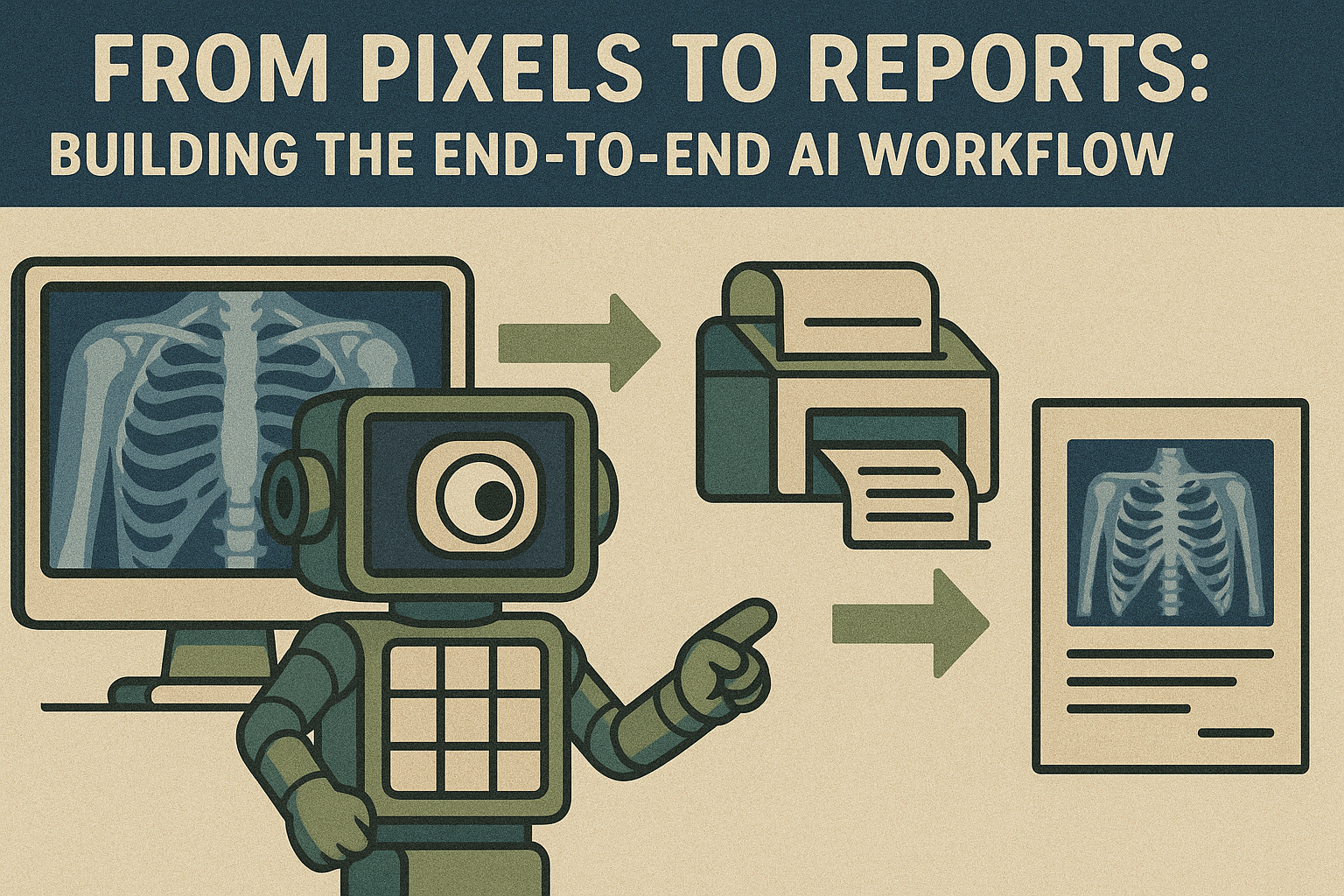
From Pixels to Reports: Building the End-to-End AI Workflow
The Journey Starts with Data Ingestion
Every AI-powered medical imaging workflow begins with raw data — typically DICOM files from modalities like X-ray, CT, MRI or ultrasound machines.
These files are rich in both images and metadata (patient details, scan parameters), but handling them safely and efficiently is crucial.
Key first steps include:
Secure upload: Images are transferred to a processing system, often through encrypted channels to meet privacy regulations like HIPAA.
De-identification: Patient names, IDs and other sensitive details are removed or masked. Tools like an Image Anonymization API help automate this process while keeping the medical information intact.
By properly preparing the data, healthcare providers can ensure privacy, security and compliance from the very beginning.
Preprocessing: Preparing Images for Analysis
Before AI models can read an image, some preparation is usually needed:
Normalization: Adjusting brightness and contrast to standard levels across different devices.
Cropping and resizing: Focusing on the regions of interest and making image sizes consistent.
Noise reduction: Cleaning up artifacts that might confuse the model.
In many cases, APIs such as Background Removal can help eliminate irrelevant parts of a scan, making it easier for the AI to focus on important areas.
Preprocessing not only improves model performance but also speeds up the entire workflow by reducing unnecessary data.
The Core: Running AI Models in Parallel
Once images are cleaned and ready, the real AI magic happens.
Different types of models often work together, each specializing in a specific task:
Detection models: Locate and highlight anomalies, such as tumors, fractures or nodules.
Segmentation models: Outline the exact shape and size of abnormalities, crucial for planning treatments like surgery or radiation.
Classification models: Categorize findings into types (benign vs malignant, normal vs abnormal).
OCR models: Read any embedded text in the image (such as labels or calibration markers).
Modern cloud systems allow these models to run in parallel, dramatically reducing the total processing time.
For example, an object detection model might identify suspicious lung nodules, while a segmentation model outlines their borders and an OCR model checks for important technical notes — all at once.
Some clinics use ready-made Object Detection and Image Labelling APIs to quickly add this intelligence into their workflows without needing to train models themselves.
Confidence Scoring and Human-in-the-Loop Review
AI is powerful but not perfect. To build trust and ensure safety, many workflows incorporate a human-in-the-loopsystem:
Each AI result is assigned a confidence score indicating how sure the model is about its finding.
Cases with lower confidence can be automatically flagged for manual review by a radiologist.
Visualization tools like heatmaps show where the AI "looked" when making its decision, offering transparency.
This approach combines the speed of automation with the judgment of human experts, ensuring that critical decisions are double-checked.
Creating Structured, Ready-to-Use Reports
Instead of giving doctors raw images with AI annotations, the best workflows generate structured reports that are easy to interpret and act upon.
A typical AI-generated report might include:
A list of detected findings (e.g., "2 pulmonary nodules detected, size: 8mm and 12mm").
Visual overlays highlighting anomalies on the original images.
Suggested next steps based on clinical guidelines.
Some advanced systems even use natural language generation to turn findings into human-readable summaries, saving doctors valuable time during busy shifts.
Integration into Existing Healthcare Systems
To be truly useful, AI outputs must flow smoothly into the systems healthcare providers already use, such as:
PACS (Picture Archiving and Communication Systems): For viewing and archiving medical images.
EHR (Electronic Health Record) systems: For storing reports and patient histories.
Hospital workflows: For case triage, billing and treatment planning.
APIs that support healthcare standards like FHIR and HL7 make this integration easier, enabling plug-and-play capabilities without major IT overhauls.
Some lightweight deployments even use mobile apps or edge devices to bring AI assistance directly to bedside care or remote clinics.
KPIs That Matter: Measuring Success
An AI imaging workflow should not just be "cool" — it must deliver real value. Key performance indicators (KPIs) include:
Sensitivity and specificity: How accurately the AI detects true positives and avoids false alarms.
Time to result: How much faster reports are generated compared to traditional methods.
Cost savings: Reduction in repeat scans, shorter hospital stays and lower radiologist overtime.
Adoption rates: How easily radiologists and doctors adapt to using AI in their daily routines.
Tracking these metrics helps hospitals refine their workflows, optimize AI performance and demonstrate the business value of their investment.
Next, we’ll explore the critical topics of deployment, compliance and building trust — essential ingredients for successfully bringing AI into clinical practice.
Deployment, Compliance and Trust
Moving from Prototype to Clinical Reality
Building an AI model that works in a lab setting is one thing. Getting that model safely and reliably into a real hospital environment is a much bigger challenge.
Deployment in healthcare means dealing with strict rules, complex systems and very high expectations for reliability.
Before AI can assist real doctors with real patients, it must pass through a careful process of integration, validation and trust-building.
Privacy and Security: Protecting Patient Data
One of the biggest concerns in medical imaging AI is data privacy.
Medical scans are deeply personal and protected by regulations like HIPAA in the United States and GDPR in Europe. To deploy AI responsibly, healthcare providers must ensure:
Encryption of data in transit (when moving between systems) and at rest (when stored).
Access controls to ensure only authorized users can view or modify sensitive information.
Audit trails that log who accessed what data and when.
Cloud-based APIs and processing platforms must also be designed with strong security measures.
For example, when using cloud services for image analysis, features like de-identification (automatically removing patient information) are critical. API4AI's Image Anonymization API is an example of a tool that can help meet these privacy requirements.
Maintaining patient trust is non-negotiable — security and transparency must be built into every step.
Regulatory Validation: Proving Safety and Effectiveness
Healthcare is one of the most tightly regulated industries in the world — and for good reason.
Before an AI model can be widely adopted, it often needs to go through a regulatory review process to prove:
Clinical safety: The model does not cause harm through false positives or negatives.
Effectiveness: The model actually improves diagnostic accuracy or workflow efficiency.
Consistency: It works reliably across different hospitals, devices and patient populations.
In the U.S., this might mean obtaining FDA clearance. In Europe, it could mean CE certification.
Achieving regulatory approval often requires extensive validation studies, including:
Multi-site testing: Verifying performance across different hospitals and regions.
Cross-scanner validation: Ensuring the AI works across various machine models and manufacturers.
Phantom studies: Using simulated datasets to test model behavior under controlled conditions.
Without this rigorous validation, AI solutions risk being seen as "nice to have" tools instead of trusted medical devices.
Continuous Monitoring: AI Must Keep Learning
Once deployed, an AI model cannot be left alone forever. Medical data can evolve — new diseases emerge, scanning technologies improve and patient populations change.
This means AI systems must include processes for:
Drift detection: Monitoring whether model performance declines over time.
Feedback loops: Allowing doctors to correct AI errors, which can be used to retrain and improve the model.
Version control: Keeping careful records of model updates and changes to ensure traceability.
Some systems even use automatic re-training based on new labeled data, creating a continuous improvement cycle that keeps the AI current and effective.
Build or Buy? Finding the Right AI Strategy
When it comes to implementing AI, healthcare organizations face a key decision:
Should they build custom models from scratch or integrate ready-made solutions?
Each path has pros and cons:
Off-the-shelf APIs offer fast, affordable ways to get started. They are ideal for common tasks like basic anomaly detection or initial triage.
Custom AI development allows healthcare providers to create highly specialized models tailored to their unique needs — for example, detecting rare diseases or analyzing highly specific imaging protocols.
Custom solutions can require more upfront investment, but over time, they may deliver greater accuracy, lower operational costs and a competitive advantage in patient care.
Providers like API4AI offer both options: ready-to-go APIs for immediate needs and custom development services for organizations that want to build strategic, future-proof AI systems.
Building Trust with Doctors and Patients
Ultimately, no AI tool will succeed without trust.
Radiologists, clinicians and patients must feel confident that AI is an ally — not a black box.
Ways to build trust include:
Explainability: Showing not just what the AI predicts, but why — for example, using heatmaps or highlight overlays.
User training: Helping doctors understand AI limitations and best practices.
Collaborative design: Involving clinicians early when developing and testing AI tools.
When doctors see AI as a transparent assistant rather than a mysterious competitor, adoption happens faster — and patients ultimately benefit from better, more timely care.
Conclusion and Next Steps
A New Era for Medical Imaging
Medical imaging has always been a cornerstone of modern healthcare. But today, thanks to deep learning, it is evolving faster than ever before.
Technologies like Convolutional Neural Networks (CNNs) and Vision Transformers (ViTs) are no longer just research experiments — they are practical tools that are already improving how doctors detect, diagnose and treat patients.
These AI models bring real benefits:
Faster analysis of complex scans.
More accurate detection of subtle abnormalities.
Better workflow efficiency, helping radiologists focus on the most urgent cases.
Higher patient safety, with fewer missed findings.
By combining the speed and consistency of AI with human clinical expertise, healthcare systems can provide better care, faster and more affordably than ever before.
AI Is a Partner, Not a Replacement
It’s important to remember that AI is not here to replace radiologists or doctors.
Instead, AI acts as a second set of eyes — helping to reduce fatigue, catch rare or hidden conditions and support clinical decision-making.
In high-stress, high-volume environments, AI can make the difference between a delayed diagnosis and a life-saving intervention.
Ultimately, AI frees up valuable time for healthcare professionals, allowing them to spend more time on complex cases, patient communication and personalized treatment planning.
Opportunities for Healthcare Providers
For hospitals, clinics and diagnostic centers, embracing AI in medical imaging is no longer optional — it’s becoming a competitive necessity.
Some practical next steps include:
Auditing current imaging workflows to identify bottlenecks where AI could help.
Piloting ready-made APIs to quickly test AI integration.
Exploring custom AI development for specialized needs, like rare disease detection or complex multi-modal imaging analysis.
Investing in training to prepare radiologists and technicians to work confidently alongside AI tools.
Starting small — with focused use cases like fracture triage or breast cancer screening — allows organizations to build experience and trust before scaling up across multiple departments.
Why Early Adoption Matters
Healthcare providers that start integrating AI now will be better positioned to:
Meet future regulatory expectations.
Attract top medical talent interested in working with cutting-edge technologies.
Deliver faster, safer and more personalized care to patients.
Gain financial advantages through operational efficiency and value-based care incentives.
Early adopters of AI-powered imaging are not just following a trend — they are shaping the future of healthcare.
Final Thoughts
The future of medical imaging is bright.
With the combined strengths of CNNs, Vision Transformers and full-stack AI workflows, healthcare is moving toward a world where diseases are caught earlier, diagnoses are made faster and treatments are delivered with greater precision.
For those ready to invest in smart, thoughtful AI integration — whether through off-the-shelf APIs or custom-built models — the rewards will be both clinical and strategic.
Medical imaging AI is not just about technology; it’s about delivering better outcomes for every patient, every time.


